PhD student Matthew Barker is working to improve a cell transplantation therapy to repair the injured spinal cord. He is in his final year of the PhD and is due to finish at the end of 2019.
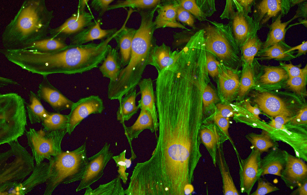
He has made a breakthrough discovery by identifying a new protein that can be used as a tool to identify different types of specialised nasal cells which are used for the transplantation. Matthew’s more recent experiments have focused on the effect that the protein has on the activity and behaviour of the cells. Using high resolution microscopy to take thousands of images and computer-based image recognition, he has observed changes in the texture of the cells’ “skeletons” in response to the proteins.
This has led him to identify the ideal conditions under which these cells grow which will improve how the cells are prepared for a successful transplantation. Matthew has also discovered that the protein can increase the rate at which the cells multiply. Increasing the rate at which the cells multiply should lead to reduced times between taking a patient’s nasal cell biopsy and transplantation into the spinal cord.

Cell pictured is a confocal microscopy image of cells taken from the nose, treated with a protein found naturally in the body, and stained with different markers of interest. Blue stains the nucleus of cells, orange stains the entire cells, and green stains the skeleton fibres (actin filaments). Understanding the changes in the actin in response to proteins may allow a better understanding of when a cell is ready for transplantation.


