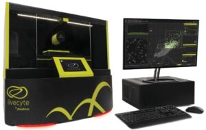
We are excited to announce that with your generous support, we have been able to fund the first piece of equipment to be used as part of the Cell Transplantation and Rehabilitation Human Clinical Trial – the PhaseFocus LiveCyte Microscope.
This incredible microscope, easily detects individual cells and provides a wealth of information about their function and behaviour. In particular, it can detect cells that have characteristics of cancer cells, such as cells that multiply and migrate rapidly. This technology therefore allows the preparation of cell transplants that contain the optimal mix of cells that stimulate regeneration (“the good cells”) whilst excluding inappropriate cells that dilute the transplant or have harmful effects (“the bad cells”).
This new analytical capability will become the gold standard in clinical cell transplantation therapies, as it will lead to cell preparations with a considerably increased safety profile. Critically, it will both optimise and de-risk the cell transplantation therapy and thereby increase the likelihood of success.
Live cell imaging is a powerful technique for the study of cultured mammalian cells, and crucial for establishing cell identity and behaviour – essential for cell transplantation. But the exposure to light and the need to label cells with chemical markers in conventional live cell imaging techniques can have toxic effects on the cells. Therefore, characterising individual cells in preparation for transplantation without impacting their health and behaviour is a significant challenge.
The Phasefocus Livecyte uses a technique called ptychographic quantitative phase imaging. This technique generates high-contrast, detailed images of live cells using low powered illumination instead of harsh light, and does not require chemical labelling of the cells. The Livecyte is also equipped with a fully enclosed temperature-controlled incubation unit with a controller for carbon dioxide and humidity to ensure that the cells remain stable, viable and healthy over the imaging period (days-weeks). Thus, it is much gentler on the cells than any other live cell microscopy technique.
The Livecyte will help us overcome the challenge of characterising cells before transplantation without causing damage, as it allows label-free and non-invasive long-term imaging. In addition, critical attributes of the cells such as individual cell behaviour, morphology and migration capacity over time can be monitored with high specificity using the Livecyte. Cells derived from tissues are a mixture of different cell types.
Tracking individual cells will help us determine the exact cell composition and heterogeneity of the cell population. The Livecyte system has automated single-cell tracking algorithms with an integrated analysis suite making it easy to measure the motion and morphology of every cell. Specific cell sub- populations can be identified, and we can stratify groups of cells from a complex cell culture based on their behaviour. Then we can generate a unique fingerprint for each cell and a comprehensive profile of its dynamics within the entire cell population. This information will improve our understanding of how each cell behaves within the environment of the actual transplant and will be a realistic measure of cell characteristics.
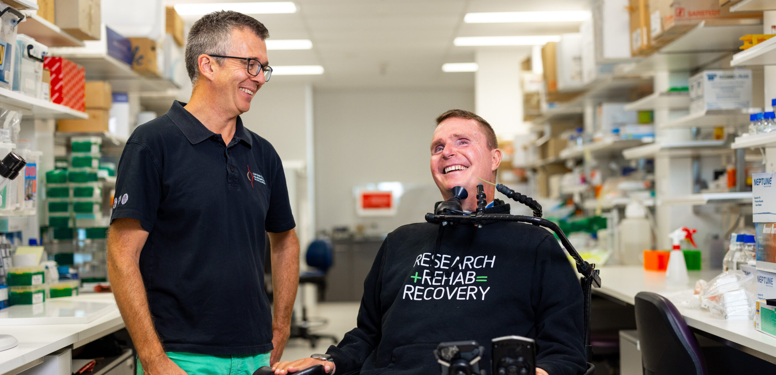
With your support we have invested $400,000 to purchase this incredible microscope so the Spinal Injury Research Team can get started!
Thank you to everyone who has donated in support of this life changing research – we are so grateful!


